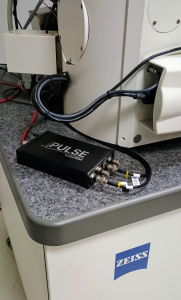
Why Pulse?
 Currently, in many technology areas such as television, car radios or photography, the switch from analog to digital has delivered better performance and lower noise. Imaging in the scanning transmission electron microscopes (SEM) should be no different, but decades since its inception, analog detectors are still commonplace.
However, although these analog detectors are still commonly installed in nearly every SEM, these are actually highly sensitive sensors and are capable of detecting single electron signals [1]. Often this sensitivity is lost when acquisition systems perform simple integration on slow timescales. Recently, research has shown that if the signal is streamed at far faster sampling rates, and if the signal is process to count each electron event rather than integrating, a new digital imaging mode is obtained [2].
The Pulse signal processing unit from turboTEM performs precisely that function, streaming the raw signal from existing analog STEM detectors at high-speed and performing edge-detection based electron counting. The digital signal is then passed out to the existing data acquisition system on the microscope. By adding the ‘Pulse’ unit to existing systems, users can experience an upgrade to digital imaging with minimal additional hardware outlay.
The benefits of this new digital imaging mode are most obvious in two scenarios, fast scanning and low-dose. When imaging at high frame-rates such as those used for in-situ imaging (e.g. dwell times shorter than 2μs), individual afterglow streaks from each electron impact can begin to smear across several image pixels degrading spatial resolution. Alternatively, when imaging at very low doses, where single electron scattering is visible in each frame, the overall image quality can be dominated by Gaussian (thermal) noise from the microscope’s readout electronics. The Pulse system overcomes this by outputting a purely digital signal, with no thermal noise. This delivers, for example in dark-field STEM, a perfect zero signal in vacuum regions.
Frequently Asked Questions
Can Pulse be used with scanning electron microscopes (SEM) and helium ion microscopes (HIM)?
Yes! Pulse works equally well on detectors on both SEM and HIM instruments.
How many channels does the Pulse read-out module support?
Pulse is currently available in either a 2-channel or a 4-channel version. The 2-channel version can be used, for example, for secondary electron (SE) and monolithic backscattered (BS) detectors. The 4-channel version can be configured to support quadrant backscatter for topographic measurement.
What scan-controllers / acquisition systems does the Pulse system work with?
The digital signals are transmitted using an industry-standard TTL digital line. This means that the Pulse unit is compatible with any acquisition system that is capable of recording digital ‘ones’. The Pulse system has been tested using the Gatan Digiscan 2* and pointElectronic ‘DISS6’ acquisition systems. The Pulse unit will also be forward compatible with any counting image acquisition system such as the Gatan DigiScan 3*.
* DigiScan is a trademark of Gatan Inc.
How is the Pulse unit connected to the Microscope?
The Pulse unit is connected to the analog outputs from the instrument’s existing detectors. This is most easily done by T-ing into the BNC connectors between these detectors and the acquisition unit (e.g. Digiscan). As the signal is read via such a “T”, the existing analog images still appear on the microscope as before and may be collected fully simultaneously with the new digital images.
[1] “Electron-count imaging in SEM“, Yamada et al., Scanning (1991).
[2] Mullarkey et al., Microscopy & Microanalysis 27 (2020).


